Subdural hematoma (SDH) is a collection of blood between the inner layer of the dura and the brain that occurs mostly as a result of trauma. It is one of the most common neurosurgical conditions in the United States today. SDH monitoring for hematoma expansion would be beneficial to enable expeditious surgical intervention. Existing imaging technologies such as MRI and CT Scans, provide excellent brain imaging information, yet fail to provide continuous monitoring for real-time hemorrhage expansion.
Novel monitoring technology developed at the Weizmann Institute of Science, incorporates a combination of hybrid radio frequency and optical (RFO) modalities into a wearable device, for continuous subdural hematoma monitoring. This device is a safe, non-invasive and easy-to-use tool, that provides precise and continuous SDH surveillance. In addition to continuous monitoring, it has the capability to alert the patient and provide real-time information to the healthcare team.
The incidence of subdural hematoma (SDH) is on a constant rise due to various factors, including aging population and increased use of blood-thinners and antiplatelet agents. Without continuous monitoring technologies to track and alert on the expansion of a SDH in real-time, patients are at a higher risk for subsequent complications and mortality. The lack of an adequate SDH detection and monitoring solution in the community following minor head injuries, results in patients often suffering severe functional deterioration and medical complications. Furthermore, due to the absence of satisfactory real-time imaging solutions, hospitals take extra precautionary measures, including prolonged hospitalization of patients and performance of repetitive tests resulting in increased expenses to the healthcare system reaching billions of dollars.
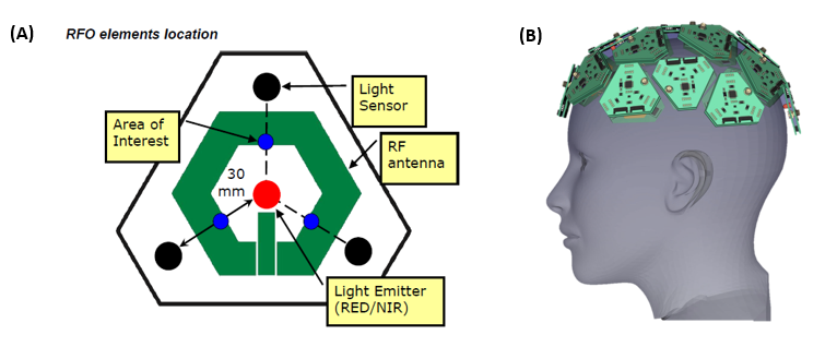
Transcranial Optical Vascular Assessment (TOVA) is a wearable device that assesses subdural cerebral blood accumulation through the intact cranium by using a combination of radio frequency and optical (RFO) mapping technologies. The optical modality is based on the synergistic effect between diffused spectroscopy sensing brain metabolism with laser speckle fluctuations detecting areas of fluid flow. The information acquired by the optical modality is processed by the dedicated AI algorithm in order to improve information recovery from the optical sensors of the device. A radiofrequency (RF) module integrated within the same sensor, assesses the hematoma by measuring the difference in the dielectric properties of blood compared to the surrounding tissues. The data obtained from each modality provides valuable information; however, their synergistic properties enhance the overall performance improving the penetration depth, spatial and temporal resolution.

Sensor design and assembly. (A) A schematic representation of the integration of an RF antenna and optic electrode (RFO) elements in a single sensor: red: light emitter, black: light sensors and green: an RF antenna. Blue: the matching areas on the skull that are at the optimal focus point of the combined modalities (areas of interest). The final design will include multiple sensors. (B) Arrangement of multiple sensors on a single device.
Advantages
- Enables continuous monitoring
- Detects and alerts on hematoma expansion in real-time
- Simple, low-cost production
- Simple operation, with no specialized training required
- Automatic, real-time results
- Detects hematomas as small as 1cm in size
Applications
- Detection and monitoring of subdural hematomas:
- Continuous monitoring during hospital admission and subsequent supervision
- Subdural and epidural assessment in ambulances and urgent care centers
- Detection of CSDHs in nursing homes and community clinics
- Long-term monitoring in community physician offices
- Identification of brain asymmetry and intracranial pressure
The technology was successfully tested in mice and on human head phantoms, using a single RFO element. Work on a lab-stage prototype is currently in progress and further human head phantom and RFO sensor optimization experiments are planned for the near future. This invention is covered by three separate patents.
Approximately 2.8 million cases of traumatic brain disorders reach the emergency room annually in the United States alone, one-third of which are SDHs at different degrees of severity. SDH accounts for approximately 4.6 million admission days, costing the healthcare system and insurance companies approximately $23 billion annually. This device will dramatically revolutionize SDH treatment by providing real-time monitoring and alerts for hematoma expansion. It will enable the detection of chronic SDHs at an early stage and reduce admission costs by enabling continuous monitoring of patients from home. It is therefore expected to be rapidly integrated into the market as a new standard-of-care for SDH.


