Cardiac fibrosis, a scarring process that impairs cardiac function and increases the risk of sudden death, remains difficult to detect at early stages with current imaging tools. This technology introduces a novel magnetization-transfer (MT) MRI method for high-sensitivity detection of myocardial fibrosis, enabling quantitative, contrast-free imaging of tissue scarring. The method generates three-dimensional fibrosis maps for diagnosis, disease monitoring, and surgical planning.
- Early and non-invasive detection of myocardial fibrosis in hypertensive, diabetic, or post-infarction patients
- Assessment of anti-fibrotic therapy efficacy and disease progression
- Surgical planning (e.g., RF ablation, cardiac assist device placement) based on 3D fibrosis geometry
- Detection of fibrosis in other organs — liver, kidney, skeletal muscle, or fibrotic cancers
- Pre-clinical platform for studying fibrosis and developing targeted therapies
- No contrast agent required
- Enhanced sensitivity versus current gadolinium-based MRI
- Compatible with existing MRI systems
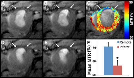
CardioCEST imaging of fibrotic scar tissue 21 days post-MI shows reduced signal and MTR in fibrotic scar regions (white arrows) compared with healthy myocardium (A, B), with persistent signal loss and absent contraction at end-systole (D, E) (*P < 0.05).
Validated in vivo in mouse models of myocardial infarction, demonstrating accurate detection and temporal monitoring of collagenous scar formation in close correlation with histology.
Pulse sequence design and analysis algorithms are established and ready for adaptation to clinical MRI systems.